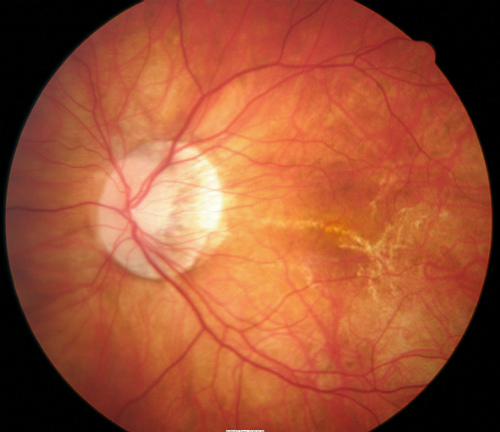

Immobile with movement of eye and produces an absolute field defect.Characteristics: Lesion is a bullous elevation of peripheral retina, most commonly found in the inferotemporal quadrant.Prevalence: found in 2-7% of the general population, more common in patients 40 years and older and in hyperopic eyes.Definition: senile retinoschisis (SR) is the splitting of layers of the neurosensory retina by thick fluid.Intra-Retinal Degenerations Senile Retinoschisis Therefore, OCT should be utilized in the initial diagnosis, treatment, and follow-up for peripheral retinal degenerations. Peripheral OCT may further aid in differentiation between types of peripheral retinal degeneration as well associated complications including subclinical retinal detachment, retinoschisis, retinal tears and vitreoretinal adhesions or tractions. Such detail includes the structure, shape and depth of retinal degenerations. Optical coherence tomography (OCT) has been shown to greatly benefit in the diagnosis of peripheral retinal degenerations by characterizing structural elements.

Chorioretinal degenerations: paving stone degeneration and peripheral retinal drusen.Vitreoretinal degenerations: lattice degeneration, snail-track degeneration, retinal tufts, and peripheral retinal breaks.Intra-retinal Degenerations: senile retinoschisis, white-without-pressure, white-with-pressure, dark-without-pressure, peripheral cystoid degeneration, snowflake degeneration, and pearl degeneration.The following are the commonly used classifications of peripheral retinal degenerations based upon depth of retinal changes observed on optical coherence tomography (OCT): Depth of retinal changes- intraretinal, retinal, vitreoretinal, or chorioretinal.Pathomorphology- trophic, tractional, atrophic, or combined.Location- equatorial, peripheral, or combined.Peripheral retinal degenerations are classified according to the following criteria: 1 Classification of Peripheral Retinal DegenerationsĬlassification of Peripheral Retinal Degenerations.


 0 kommentar(er)
